|
Physalis crassifolia |
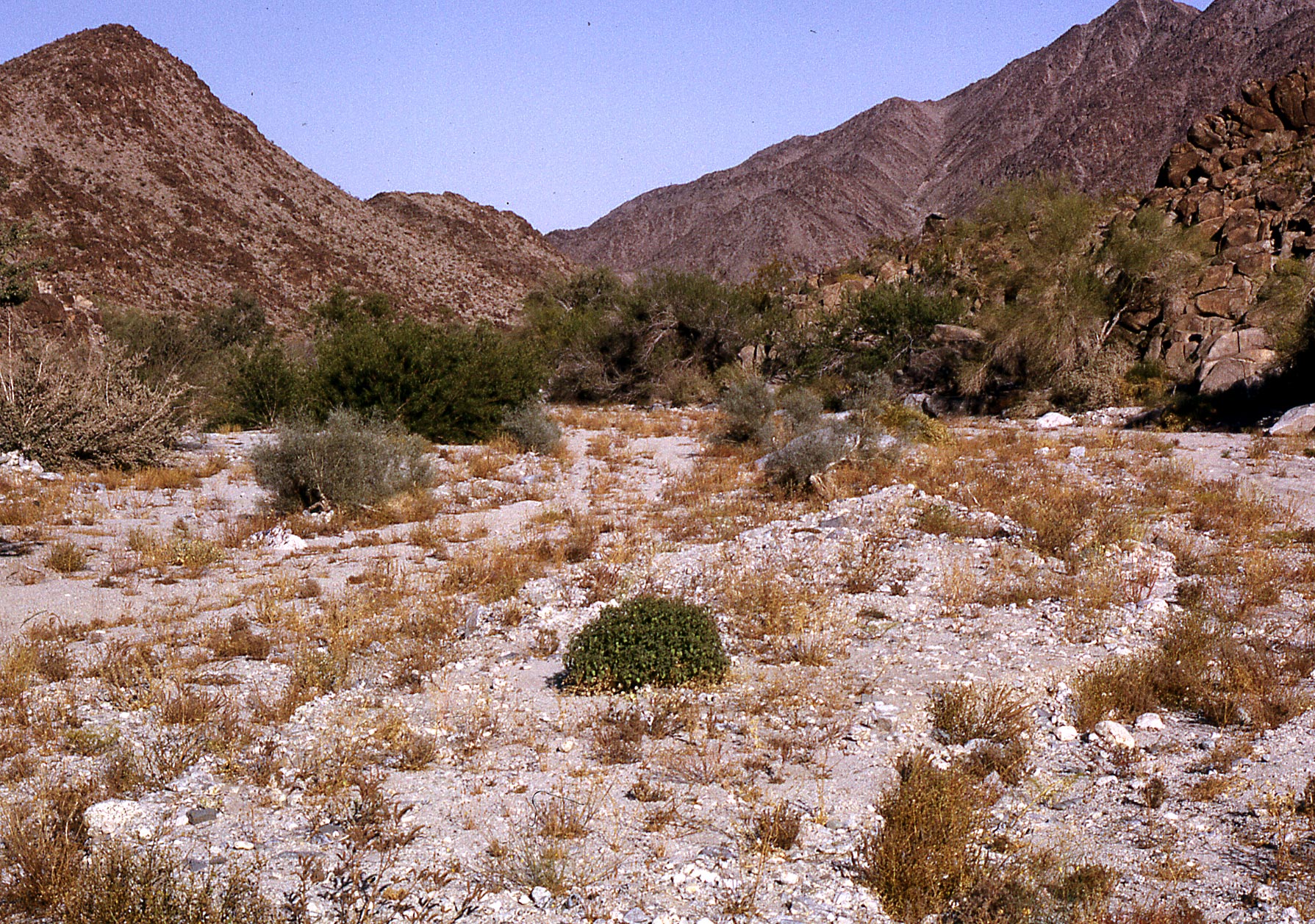
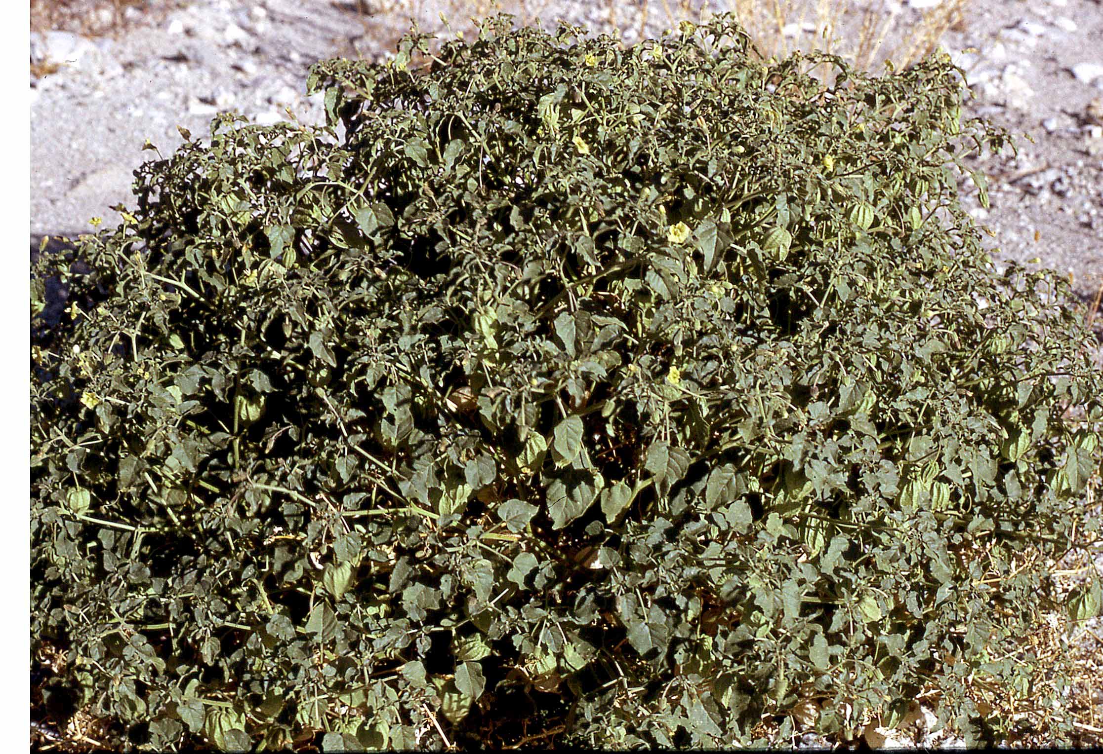
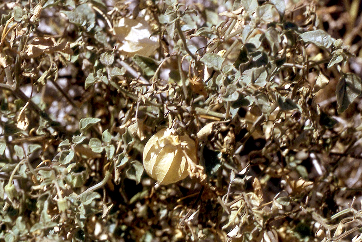
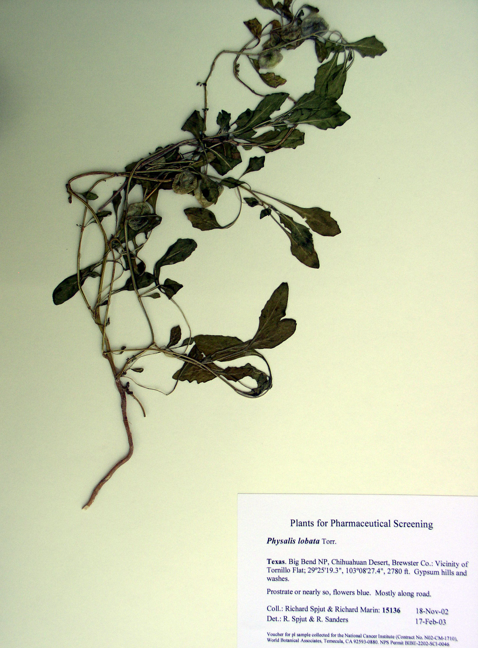
Physalis lobata
|
| Physalis maritima | |
|
Franco LA, Matiz GE, Calle J, Pinzón R, Ospina LF. 2007. [Antiinflammatory activity of extracts and fractions obtained from Physalis peruviana L. calyces] Biomedica 27(1):110–115. “INTRODUCTION: Cape gooseberry calyces (Physalis peruviana) have been used in folk medicine for their medicinal properties including anticancer, antimycobacterial, antipyretic, diuretic, immunomodulatory and antiinflammatory properties. OBJECTIVE: The antiinflammatory effect was evaluated for extracts and fractions obtained from Physalis peruviana calyces in a mice model of acute inflammation. The fractions responsible for antiinflammatory activity were extracted for possible identification. MATERIALS AND METHODS: The Physalis peruviana calyces were extracted by percolation with organic solvents. The primary hydroalcoholic fraction was purified by column chromatography. The antiinflammatory effect of extracts and fractions was evaluated using the 12-O-tetradecanoylphorbol-13-acetate-induced mouse model of ear edema. RESULTS: Thirty-eight secondary fractions were obtained by column chromatography of primary hydroalcoholic fraction. Six fractions, evaluated in 12-O-tetradecanoylphorbol-13-acetate-induced inflammation assay, showed significant antiinflammatory activity (p<0.05). The major fraction, Pp-D28-LF, showed a significant dose-dependent response at doses over 250 microg/ear. CONCLUSION: The antiinflammatory activity attributed to Physalis peruviana calyces was confirmed and validated its use in folk medicine. Fractions responsible for the antiinflammatory action were identified and seem promising for phytomedicinal development. Further studies are needed to isolate and identify the active constituents of these fractions as well as to ascertain the mechanisms involved in the antiinflammatory effect.” Hsieh W. T., K. Y. Huang, H. Y. Lin and J. G. Chung. 2006. Physalis angulata induced G2/M phase arrest in human breast cancer cells. Food Chem. Toxicol. 44(7): 974–983. “Physalis angulata (PA) is employed in herbal medicine around the world. It is used to treat diabetes, hepatitis, asthma and malaria in Taiwan. We have evaluated PA as a cancer chemopreventive agent in vitro by studying the role of PA in regulation of proliferation, cell cycle and apoptosis in human breast cancer cell lines. PA inhibited cell proliferation and induced G2/M arrest and apoptosis in human breast cancer MAD-MB 231 and MCF-7 cell lines. In this study, under treatment with various concentrations of PA in MDA-MB 231 cell line, we checked mRNA levels for cyclin A and cyclin B1 and the protein levels of cyclin A and cyclin B1, Cdc2 (cyclin-dependent kinases), p21(waf1/cip1) and P27(Kip1) (cyclin-dependent kinase inhibitors), Cdc25C, Chk2 and Wee1 kinase (cyclin-dependent kinase relative factors) in cell cycle G2/M phase. From those results, we determined that PA arrests MDA-MB 231 cells at the G2/M phase by (i) inhibiting synthesis or stability of mRNA and their downstream protein levels of cyclin A and cyclin B1, (ii) increasing p21(waf1/cip1) and P27(kip1) levels, (iii) increasing Chk2, thus causing an increase in Cdc25C phosphorylation/inactivation and inducing a decrease in Cdc2 levels and an increase in Wee1 level. According to the results obtained, PA appears to possess anticarcinogenic properties; these results suggest that the effect of PA on the levels of phosphorylated/inactivated Cdc25C are mediated by Chk2 activation, at least in part, via p21(waf1/cip1) and P27(kip1) cyclin-dependent kinase inhibitors pathway to arrest cells at G2/M phase in breast cancer carcinoma cells. Soares M. B., D. Brustolim, L. A. Santos, M. C. Bellintani, F. P. Paiva, Y. M. Ribeiro, T. C. Tomassini and R. Ribeiro Dos Santos. 2006. Physalins B, F and G, seco-steroids purified from Physalis angulata L., inhibit lymphocyte function and allogeneic transplant rejection. Int Immunopharmacol. 6(3): 408–414. “Physalis angulata is a solanaceae widely used in folk medicine in various tropical countries in the world. We have previously described that seco-steroids (physalins) purified from P. angulata are potent inhibitors of macrophage activation, blocking the production of pro-inflammatory cytokines and LPS-induced lethality. Herein we investigated the immunomodulatory activities of these substances in lymphocyte proliferation and cytokine production and in transplantation. The addition of physalins B, F or G to concanavalin A-activated splenocyte cultures induced a concentration-dependent inhibition of proliferation. Physalin B also inhibited IL-2 production by Con A-activated spleen cells. The addition of 2 mug/ml physalin B to mixed lymphocyte reaction (MLR) caused a 100% inhibition of proliferation. More importantly, treatment of mice with physalin B, F or G prevented the rejection of allogeneic heterotopic heart transplant. Our results demonstrate the suppressive activity of physalins B, F and G in lymphocyte function and indicate the potential use of physalins as immunosuppressive agents for treatments of pathologies in which inhibition of immune responses is desired. Wu S. J., L. T. Ng, D. L. Lin, S. N. Huang, S. S. Wang and C. C. Lin. 2004. Physalis peruviana extract induces apoptosis in human Hep G2 cells through CD95/CD95L system and the mitochondrial signaling transduction pathway. Cancer Lett. 215(2):199–208. “Physalis species is a popular folk medicine used for treating cancer, leukemia, hepatitis and other diseases. Studies have shown that the ethanol extract of Physalis peruviana (EEPP) inhibits growth and induces apoptotic death of human Hep G2 cells in culture, whereas proliferation of the mouse BALB/C normal liver cells was not affected. In this study, we performed detailed studies to define the molecular mechanism of EEPP-induced apoptosis in Hep G2 cells. The results further confirmed that EEPP inhibited cell proliferation in a dose- and time-dependent manner. At 50 microg/ml, EEPP significantly increased the accumulation of the sub-G1 peak (hypoploid) and the portion of apoptotic annexin V positive cells. EEPP was found to trigger apoptosis through the release of cytochrome c, Smac/DIABLO and Omi/HtrA2 from mitochondria to cytosol and consequently resulted in caspase-3 activation. Pre-treatment with a general caspase inhibitor (z-VAD-fmk) prevented cytochrome c release. After 48 h of EEPP treatment, the apoptosis of Hep G2 cells was found to associate with an elevated p53, and CD95 and CD95L proteins expression. Furthermore, a marked down-regulation of the expression of the Bcl-2, Bcl-XL and XIAP, and up-regulation of the Bax and Bad proteins were noted. Taken together, the present results suggest that EEPP-induced Hep G2 cell apoptosis was possibly mediated through the CD95/CD95L system and the mitochondrial signaling transduction pathway.”
|
|