|
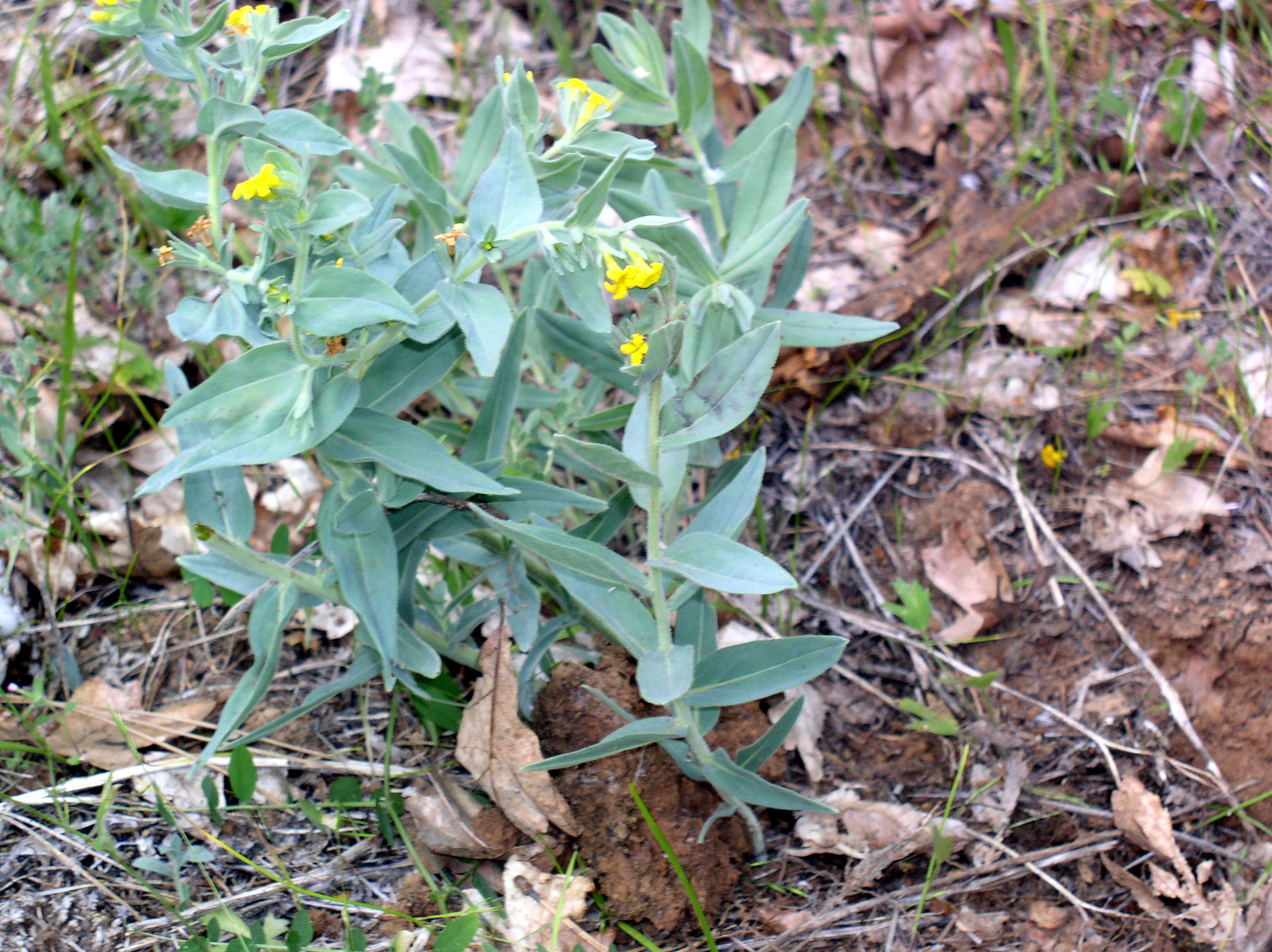
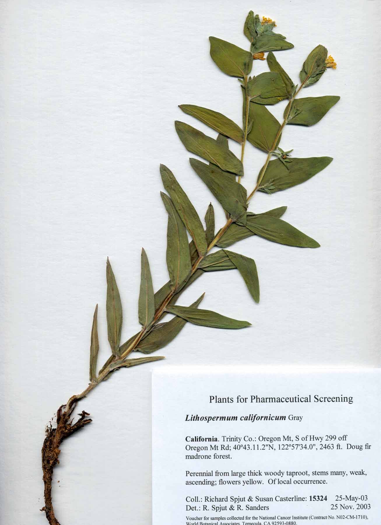
Lithospermum californicum |
Lithospermum californicum |
|
An S.,Y. D. Park, Y. K. Paik, T. S. Jeong and W. S. Lee WS. 2007. Human ACAT inhibitory effects of shikonin derivatives from Lithospermum erythrorhizon. Bioorg Med Chem Lett. 15;17(4):1112–1116. “Three naphthoquinones were isolated by bioassay-guided fractionation from the CHCl(3) extracts of roots of Lithospermum erythrorhizon. They were identified as acetylshikonin (1), isobutyrylshikonin (2), and beta-hydroxyisovalerylshikonin (3) on the basis of their spectroscopic analyses. The compounds 1-3 were tested for their inhibitory activities against human ACAT-1 (hACAT-1) or human ACAT-2 (hACAT-2). Compound 2 preferentially inhibited hACAT-2 (IC(50)=57.5microM) than hACAT-1 (32% at 120microM), whereas compounds 1 and 3 showed weak inhibitory activities in both hACAT-1 and -2. To develop more potent hACAT inhibitor, shikonin derivatives (5-11) were synthesized by semi-synthesis of shikonin (4), which was prepared by hydrolysis of 1-3. Among them, compounds 5 and 7 exhibited the strong inhibitory activities against hACAT-1 and -2. Furthermore, we demonstrated that compound 7 behaved as a potent ACAT inhibitor in not only in vitro assay system but also cell-based assay system.” Cui X. R., M. Tsukada, N, Suzuki, T. Shimamura, L. Gao, J. Koyanagi, F. Komada and S. Saito. 2007. Comparison of the cytotoxic activities of naturally occurring hydroxyanthraquinones and hydroxynaphthoquinones. Eur. J. Med. Chem. Sep. “Seven hydroxyanthraquinone derivatives, 1-7, were isolated from the root of Rheum palmatum (Polygonaceae). Two propionated anthraquinone derivatives, 8 and 9, were synthesized. Four hydroxynaphthoquinone derivatives, 13, 14, 16 and 21, were isolated from the root of Lithospermum erythrorhizon Sieb. et Zucc. (Boraginaceae) and also three naphthoquinone derivatives, 19, 22 and 23, were isolated from the root of Macrotomia euchroma (Royle) Pauls. (Boraginaceae). The cytotoxicity of the anthraquinone and naphthoquinone derivatives on P-gp-underexpressing HCT 116 cells and P-gp-overexpressing Hep G2 cells was examined by MTT assay. Among the anthraquinone derivatives, compounds 3-5 which had OH, CH(2)OH and COOH substituent groups on the anthraquinone skeletons, respectively, showed potent growth inhibitory activities against both types of cancer cells (IC(50) values: 5.7+/-0.9 to 13.0+/-0.7muM in the case of HCT 116 cells and 5.2+/-0.7 to 12.3+/-0.9muM in the case of Hep G2 cells). All hydroxynaphthoquinone derivatives isolated in this study exhibited extremely potent growth inhibitory activities against both types of cancer cells (IC(50) values: 0.3+/-0.09 to 0.46+/-1.0muM in the case of HCT 116 cells and 0.22+/-0.03 to 0.59+/-0.06muM in the case of Hep G2 cells) as well as shikonin 10 (IC(50) values: 0.32+/-0.02muM in the case of HCT 116 cells and 0.24+/-0.03muM in the case of Hep G2 cells).” Hou Y., T. Guo, C. Wu, X. He and M. Zhao. 2006. Effect of shikonin on human breast cancer cells proliferation and apoptosis in vitro. Yakugaku Zasshi. 126(12): 1383–1386. “Shikonin, isolated from the plant Lithospermum erythrorhizon Sieb. Et Zucc, has been reported to induce apoptosis in several tumor cells. However, such effect of shikonin on human breast cancer cells has not been reported. Thus, in the present study, whether shikonin could induce MCF-7 human breast cancer cell apoptosis was investigated. The results showed that shikonin (2.5-80 microM) induced MCF-7 cell death in a time- and dose-dependent manner, as measured by MTT assay. The IC(50) of a 24 h, 48 h and 72 h time course for MCF-7 cells was 7.4+/-0.4, 6.3+/-0.6 and 3.9+/-0.5 microM, respectively. Cellular morphology observation showed that MCF-7 cells underwent marked apoptotic morphological changes upon treatment with 10 microM shikonin compared with the untreated control. Flow cytometric analysis of shikonin-treated MCF-7 cells showed that the ratio of the apoptotic DNA fragmentation increased in a dose-dependent manner. The present study demonstrated for the first time that the cytotoxic effect of shikonin on MCF-7 cells underwent apoptosis process.” Ishida T, and I. SakaguchiI. 2007. Protection of human keratinocytes from UVB-induced inflammation using root extract of Lithospermum erythrorhizon. Biol. Pharm. Bull. 30(5):928–934. “UVB irradiation is an important inducer of biological changes in skin and can activate inflammatory reactions and apoptotic pathways, leading to skin damage. A root extract of Lithospermum erythrorhizon (SK), which has naphthoquinone pigments containing shikonin and shikonin derivatives, is known for its anti-inflammatory, anti-bacterial, and anti-tumor activity, and for its scavenging of reactive oxygen species. However, the effect of SK against UV damage is not clear. The aim of this study was to evaluate the efficacy of SK against UVB induced damage in normal human epidermal keratinocytes (NHEK). UVB-irradiated NHEK showed decreased cell viability, increased production of interleukin (IL)-1alpha, IL-6, IL-8, and tumor necrosis factor-alpha, and induced apoptosis. In an apoptosis pathway assay, UVB-irradiated NHEK showed increased caspase-3 activity, p53 and its phosphorylation at serine 15 compared with non-irradiated cells. All these effects induced by UVB irradiation were clearly inhibited by treatment with SK before and after UVB irradiation for 24 h. It is suggested that SK can protect epidermal cells against harmful effects of UVB irradiation and that SK treatment is probably beneficial for photoprotection of the skin.” Kim E. K., E. Y. Kim, P. D. Moon, J. Y. Um, H. M. Kim, H. S. Lee, Y. Sohn, S. K. Park, H. S. Jung and N. W. Sohn. 2007. Lithospermi radix extract inhibits histamine release and production of inflammatory cytokine in mast cells. Biosci. Biotechnol. Biochem. 71(12): 2886–2892. “Lithospermi radix (LR, Borraginaceae, the root of Lithospermum erythrorhizon Siebold. et Zuccarinii, is used in herbal medicine to treat such conditions as eczema, skin burns and frostbite. This study investigates the effects of LR on the anti-allergy mechanism. LR inhibited the release of histamine from rat peritoneal mast cells by compound 48/80 in a dose-dependent manner. LR orally administered at 6.59 mg/100 g also inhibited the anti-DNP IgE-induced passive cutaneous anaphylaxis reaction. LR inhibited the PMA plus A23187-induced increase in IL-6, IL-8, and TNF-alpha expression in HMC-1 cells. In addition, LR also inhibited nuclear factor-kappa B (NF-kappaB) activation and I kappaB-alpha degradation. These results show that LR had an inhibitory effect on the atopic allergic reaction. Furthermore, the in vivo and in vitro anti-allergic effect of LR suggests possible therapeutic applications of this agent for inflammatory allergic diseases.”
|
|