|
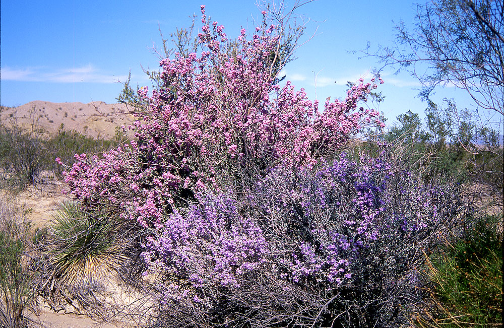
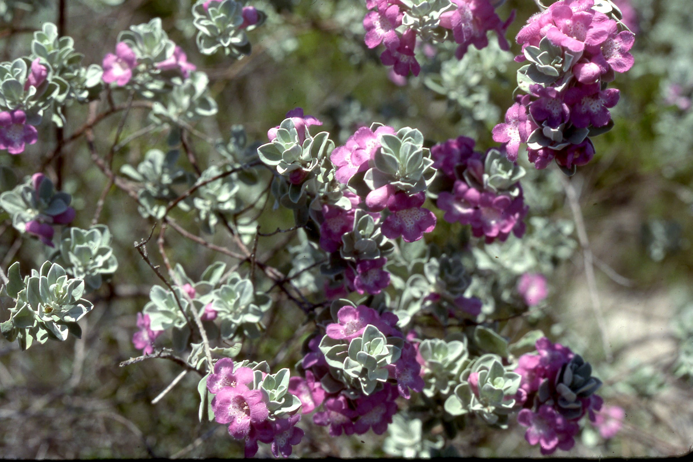
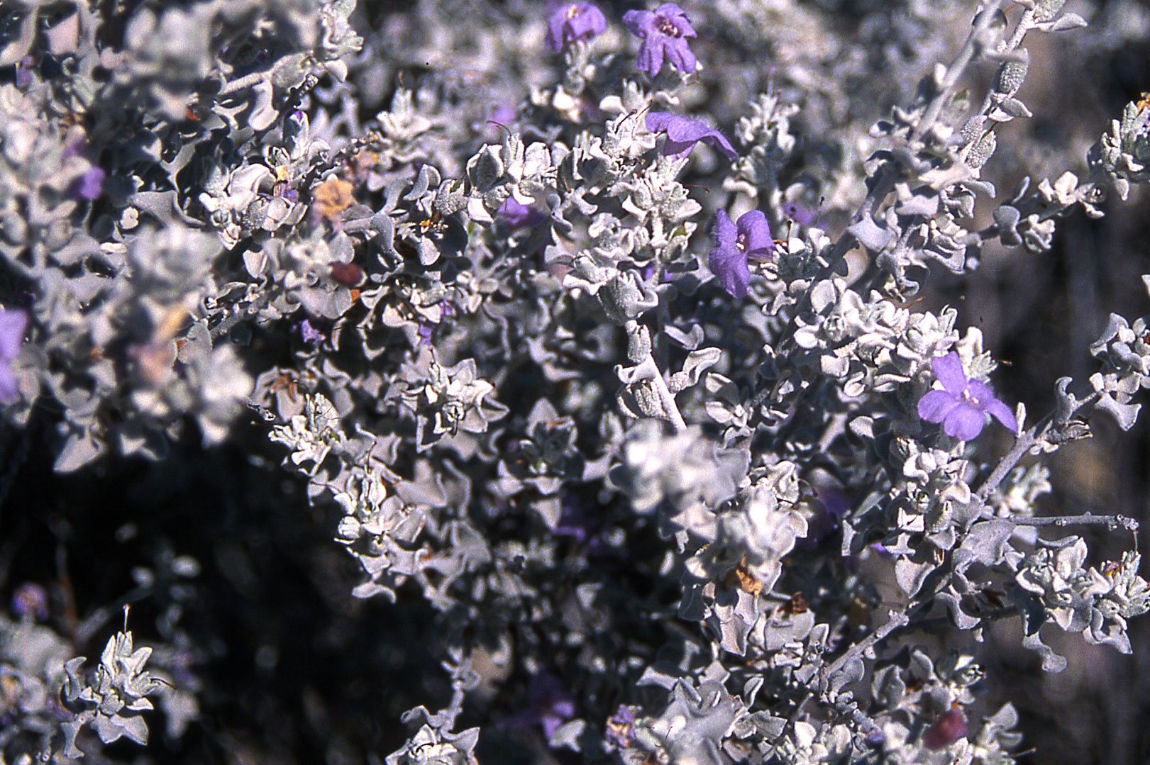
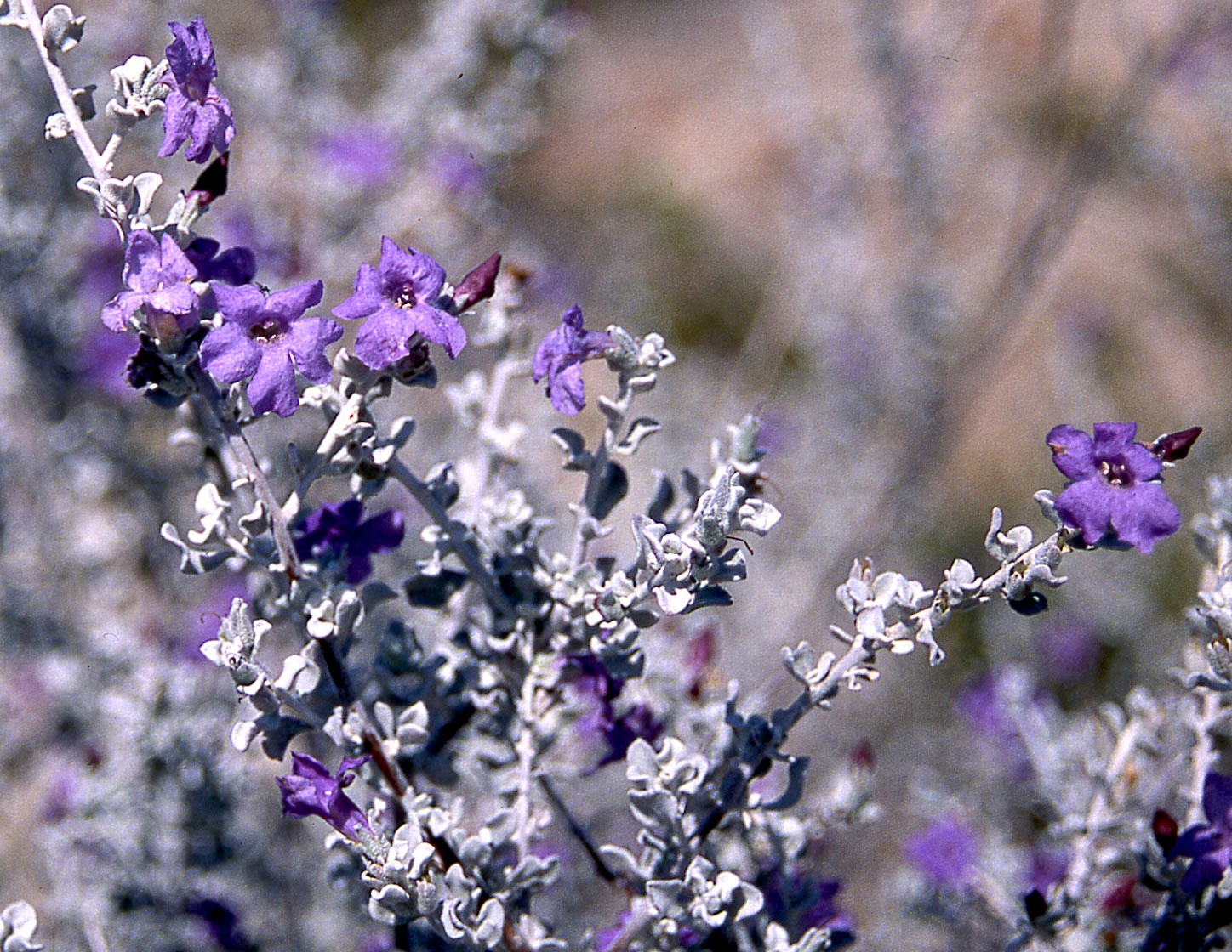
Leucophyllum frutescens and Leucophyllum candidum Big Bend National Park, TX. Sierra Del Carmen near Roys Peak, Sep 2001
|
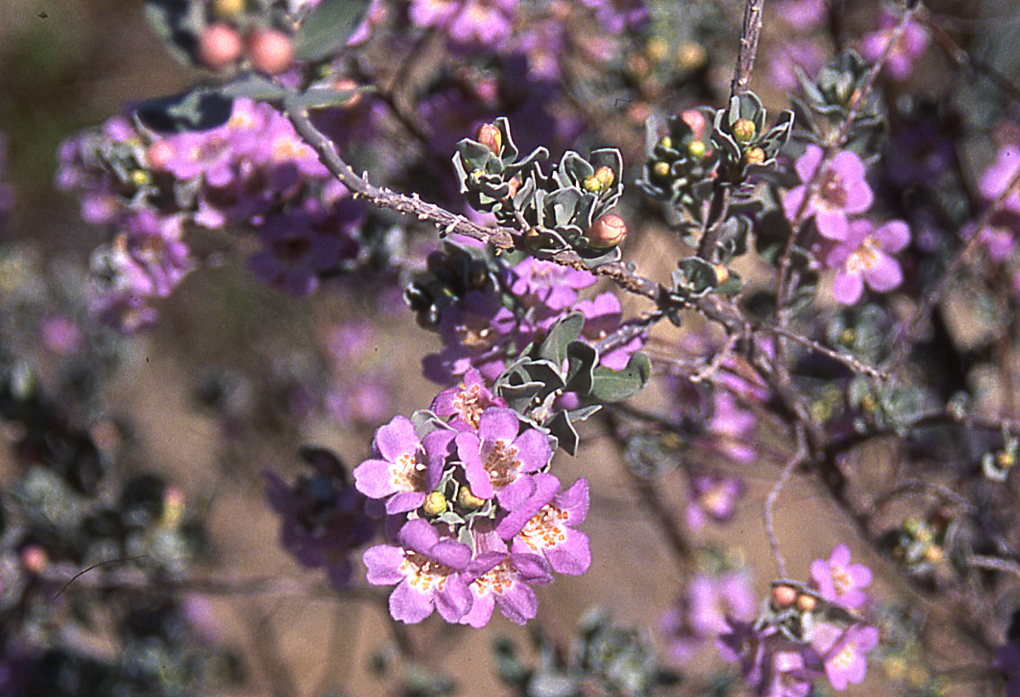
Leucophyllum frutescens
Big Bend National Park, TX. Sierra Del
Carmen near Roys Peak
|
|
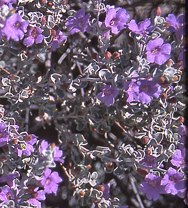
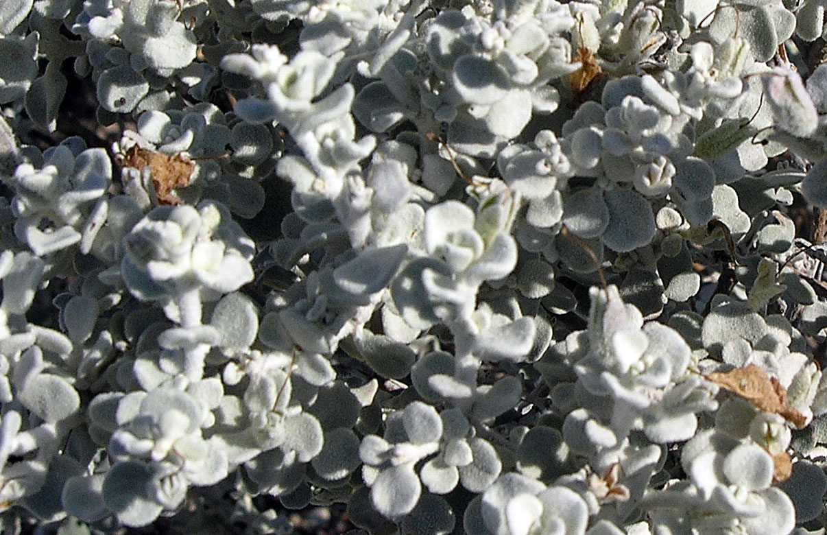
Leucophyllum
candidum
|
   Leucophyllum
candidum
|
|
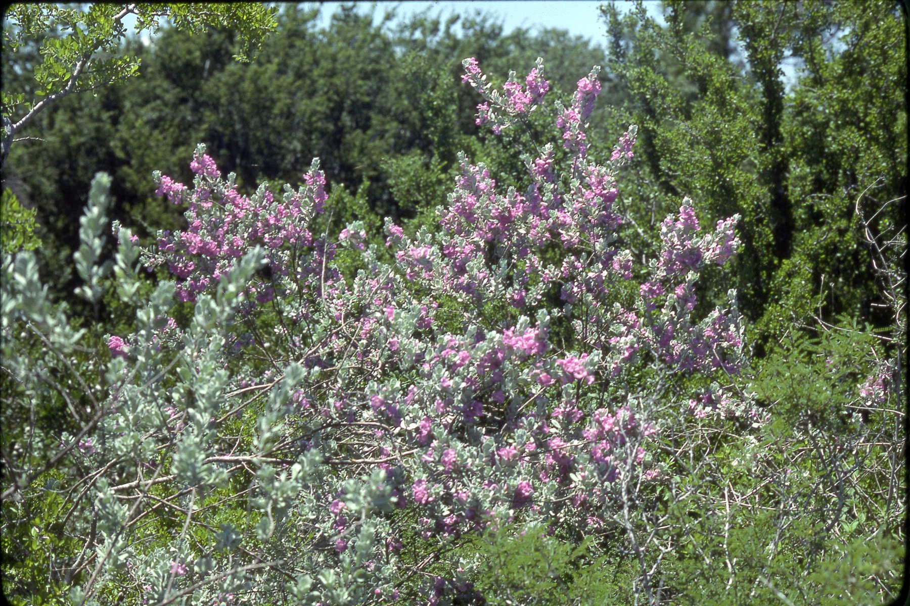
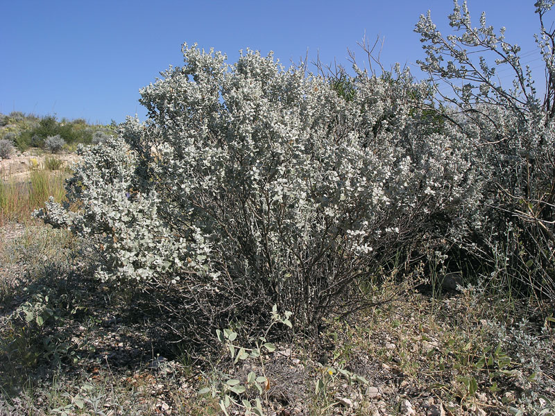
Leucophyllum frutescens
|
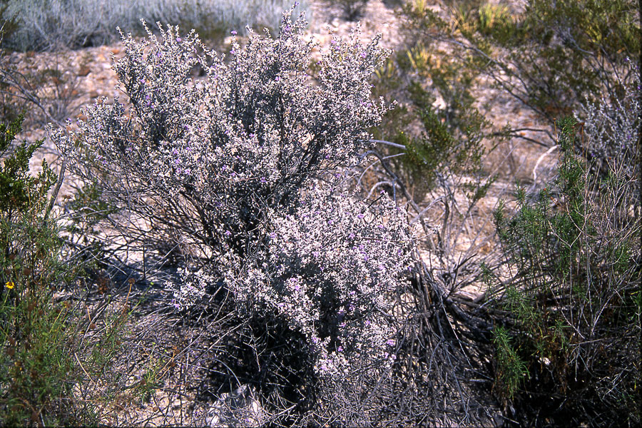
Leucophyllum minus
|
|
Balderas-Renteria I., R. Camacho-Corona Mdel, P. Carranza-Rosales, H. G. Lozano-Garza, D. Castillo-Nava, F. J. Alvarez-Mendoza and E. M. Tamez-Cantú. 2007. Hepatoprotective effect of Leucophyllum frutescens on Wistar albino rats intoxicated with carbon tetrachloride. Ann. Hepatol. 6(4): 251–254. “Many hepatoprotective herbal preparations have been recommended in alternative systems of medicine for the treatment of hepatic disorders. No systematic study has been done on protective efficacy of Leucophyllum frutescens to treat hepatic diseases. Protective action of L. frutescens methanol extract (obtained by maceration) was evaluated in an animal model of hepatotoxicity induced by carbon tetrachloride (CCl(4)). Wistar albino rats were divided into five groups. Group I was normal control group; Groups II-V received CCl(4). After inducing hepatic damage, Group II served as control CCl(4); Group III was given silymarin as reference hepatoprotective; and Groups IV and V received different doses of plant extract. Liver marker enzymes were assayed in serum. Samples of livers were observed under microscope for the histopathological changes. Levels of marker enzymes such as alanine aminotransferase (ALT) and aspartate aminotransferase (AST) were increased significantly in CCl(4) treated rats (Group II). Groups IV and V intoxicated with CCl(4) and treated with L. frutescens methanol extract significant decreased the activities of these two enzymes. Also these groups resulted in less pronounced destruction of the liver architecture, there is not fibrosis and have moderate inflammation compared with Group II. The present study scientifically validated the traditional use of L. frutescens for liver disorders. In conclusion the methanol extract of L. frutescens aerial parts could be an important source of hepatoprotective compounds. Domínguez X. A. and A. Raigosa. 1969. Chemical components of Leucophyllum texanum. Isolation of regiomontane. Planta Med. 17(4): 366–368. Rimando A. M., F. E. Dayan, J. R. Mikell and R. M. Moraes. 1999. Phytotoxic lignans of Leucophyllum frutescens. Nat. Toxins 7(1): 39–43. “Bioassay-guided fractionation of the hexane:ethyl acetate (1:1) extract of the leaves of Leucophyllum frutescens (Berl.) I.M.Johnst (Scrophulariaceae) led to the isolation of its phytotoxic constituents diayangambin (1), epiyangambin (2), diasesartemin (3) and epiashantin (4). Phytotoxicity was demonstrated as inhibition of seed germination of Agrostis stolonifera cv. penncross (Poaceae) and inhibition of development of Lactuca sativa L. (Asteraceae) seedlings in a microassay using 24-well plates. Compound 1 was the most phytotoxic to L. sativa, showing strong inhibitory activity at 110 microM. Compound 1 was more active than 2 and 3 in inhibiting the growth of A. stolonifera with I(50) values of 160, 670 and 930 microM, respectively. At a concentration of 500 microM, these compounds inhibited all phases of onion root cell division. This is the first demonstration of antimitotic activity of these furofuran lignans, and the first report of their isolation from this species.”
|
|