| Astragalus agnicidus Barneby Fabaceae WBA-1214, pl 50 g (6 bags) for NCI Biological Screening, June 1990, California, rare plant; see ltr: Spjut to Snader, plants reportedly grown from seed then harvested for sample. No activity found. |
|
  Astragalus beckwithii Great Basin Desert—Nevada. Nye Co.: E of Lida, Hwy 266; 37º27'02.7", 117º30.57.7", 1938 m. Pinyon-juniper to Joshua tree woodland. SPJ-16284, 08 May 2008
|
|
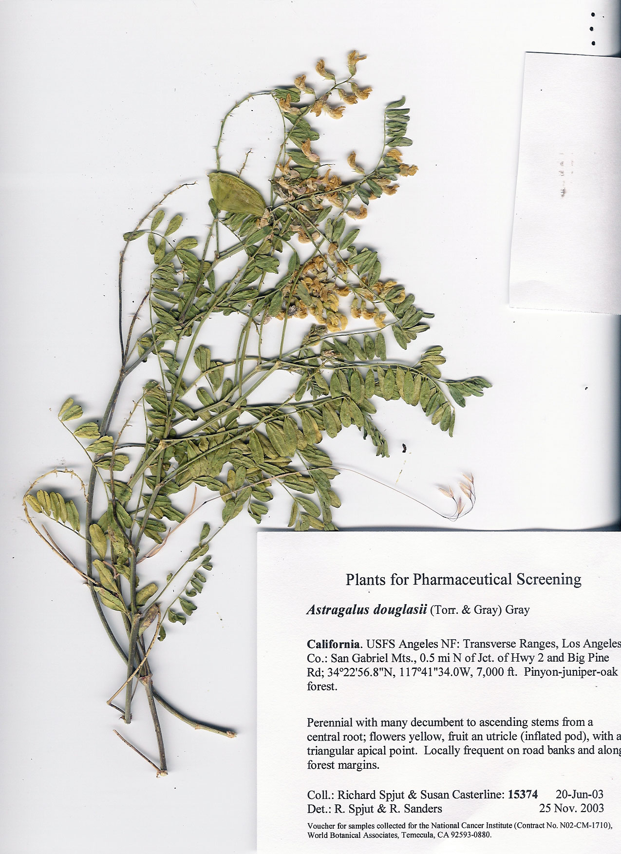
Astragalus douglasii
|
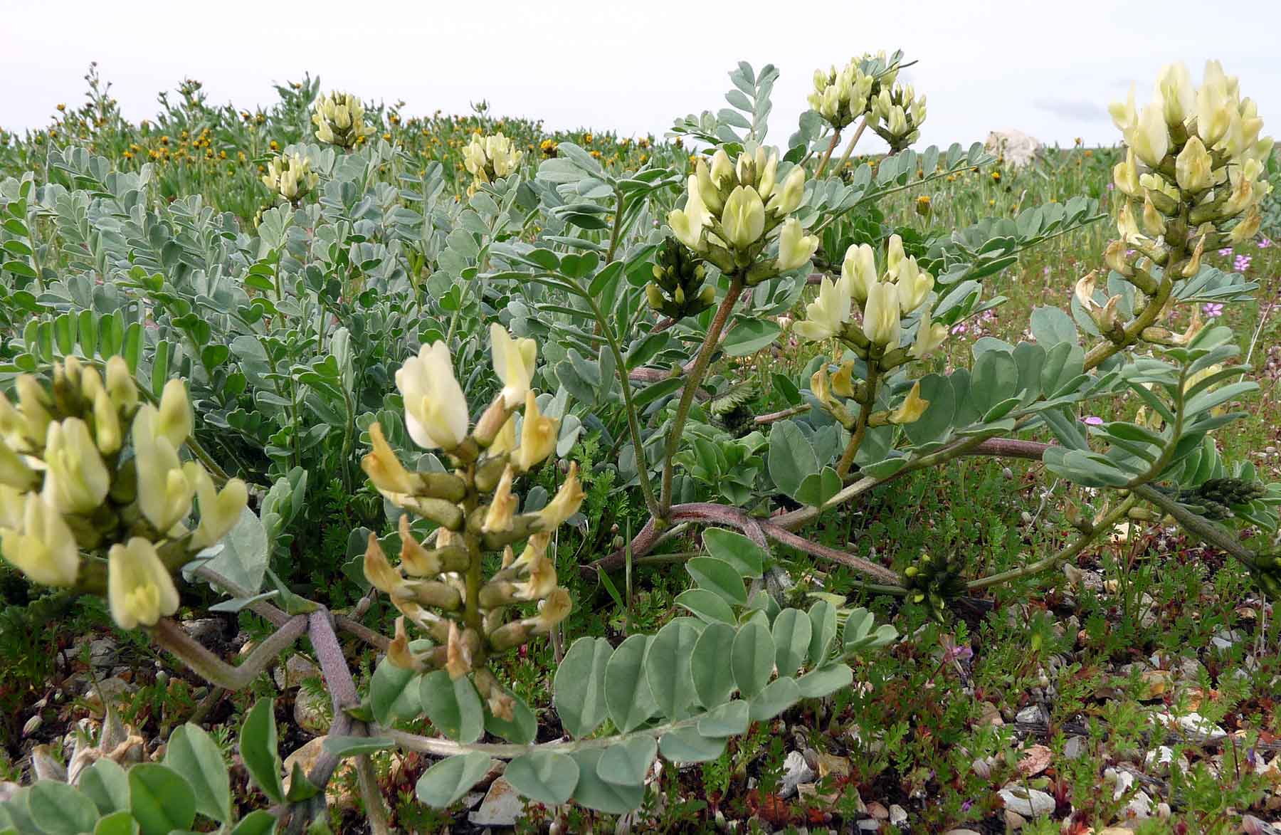
Astragalus giganteus |
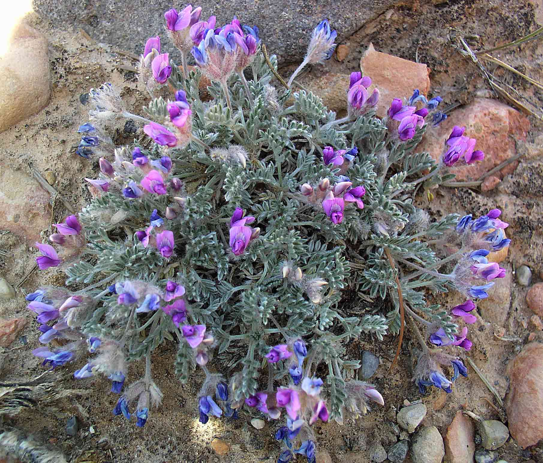
Astragalus cf insularis
|
|
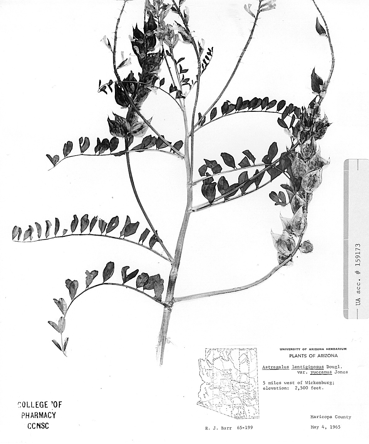
Astragalus lentiginosus AZ: 5 miles W of Wikenberg, Barr 65-199, 4 May 1965 |
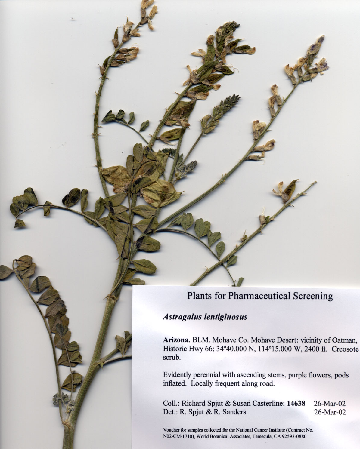
Astragalus lentiginosus
|
Astragalus lentiginosus Elkhorn Plain, CA
|
|
Astragalus lentiginosus Kern Co.: Elkhorn Plain, CA |
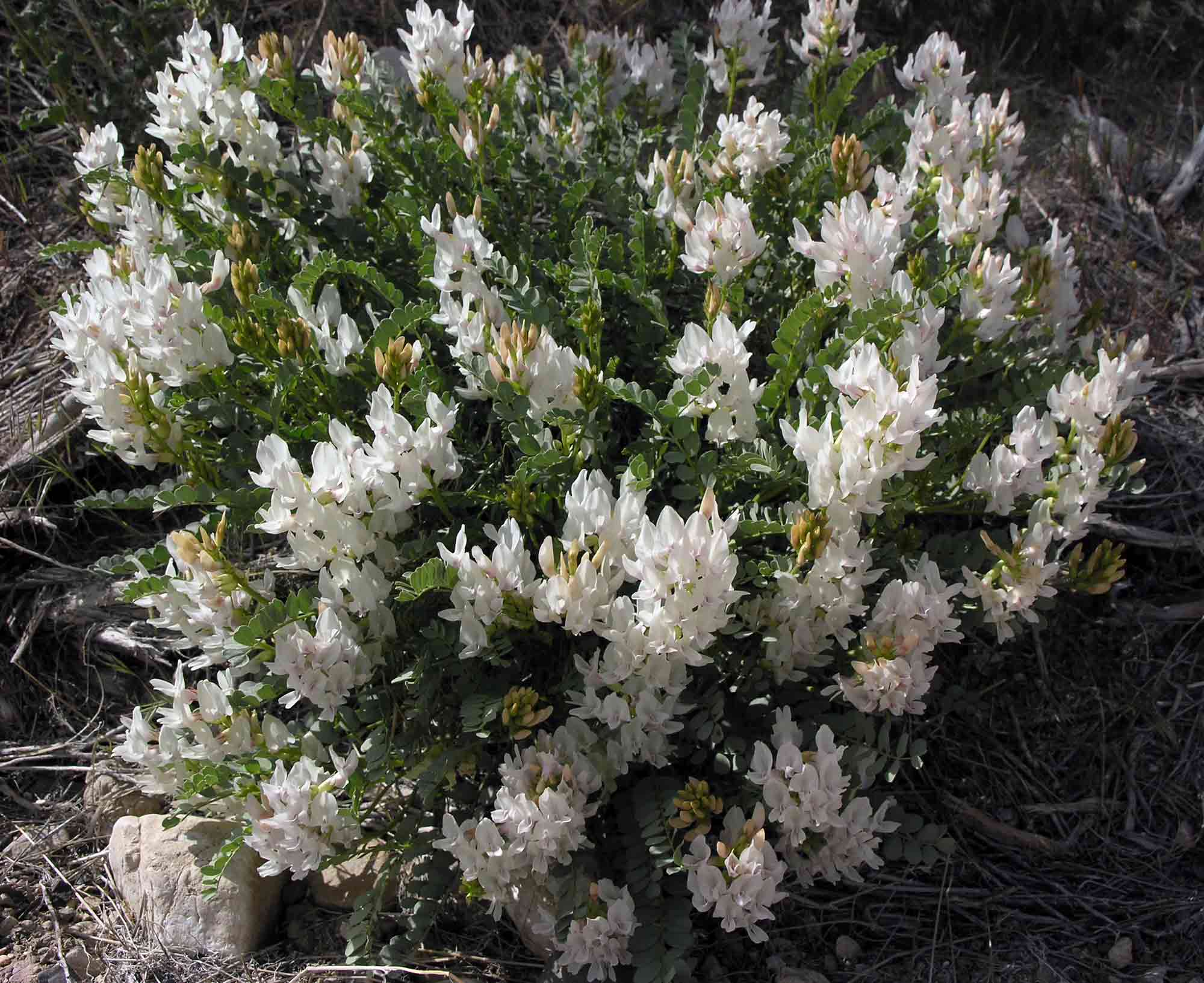
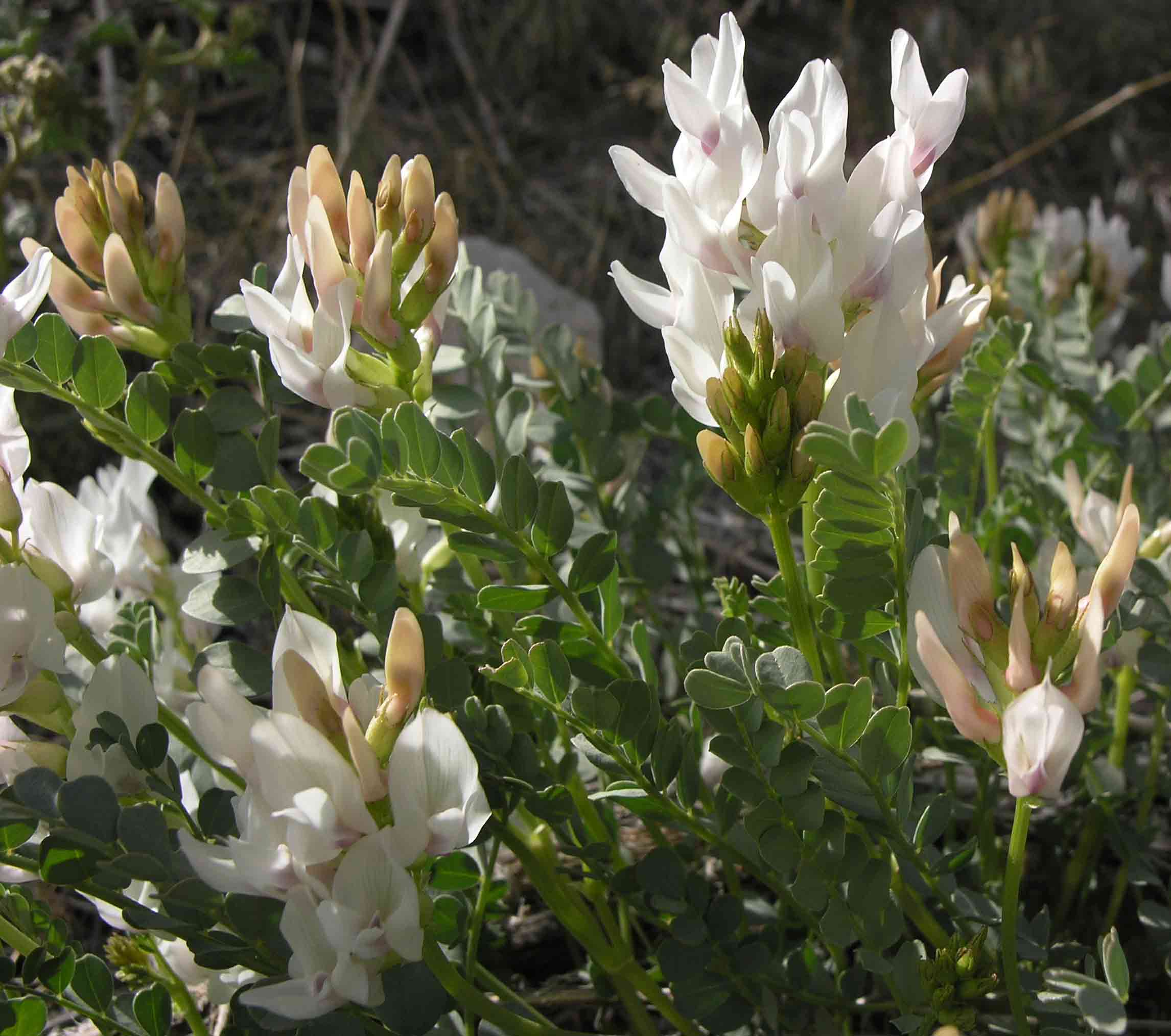
Astragalus minthorniae
|
Astragalus purshii Red Rock Canyon, UT
|
|
Abbas F., and R. Zayed. 2005. Bioactive saponins from Astragalus suberi L. growing in Yemen. Z. Naturforsch. [C] 60(11-12): 813–820. “From the aerial parts of Astragalus suberi L., Fabaceae, seven saponins were isolated. Based on spectral data (IR, 1H and 13C NMR and HR-FABMS), the structures were established as 3-O-(beta-D-glucopyranosyl)-soyasapogenol B (1); 3-O-(beta-D-glucuronopyranosyl)-soyasapogenol B (2); 3-O-[beta-D-galactopyranosyl (1-->2)-beta-D-glucopyranosyl]-soyasapogenol B (3); 3-O-[alpha-L-rhamnopyranosyl (1-->2)-beta-D-galactopyranosyl (1-->2)-beta-D-glucopyranosyl]-soyasapogenol B (4); 3-O-[beta-L-rhamnopyranosyl (1-->2)-beta-D-galactopyranosyl (1-->2)-beta-D-glucopyranosyl]-11-hydroxy-soyasapogenol B (5); 3-O-[alpha-L-rhamnopyranosyl (1-->2)-beta-D-galactopyranosyl (1-->2)-beta-D-glucuronopyranosyl]-soyasapogenol B (6) and 3-O-[alpha-L-rhamnopyranosyl (1-->2)-beta-D-galactopyranosyl (1-->2)-beta-D-glucuronopyranosyl]-complogenin (7). The isolated saponins exhibited antibacterial activities against Gram-positive and Gram-negative bacteria with minimum inhibitory concentration values >100 microg/ml, antifungal activity against all the strains tested with minimum fungicidal concentration values between 25 and 50 microg/ml and inhibited the growth of Hep-2 (human carcinoma of larynx), with IC50 values between 50 microg/ml (compounds 5-7) and 100 microg/ml (compounds 1-4), and Hela (human carcinoma of cervix) cell lines in culture with different IC50 values [74 (compound 7), 98 (compound 5) and 180 microg/ml (compounds 1-4 and 6)].” Chen XJ, Z. P. Bian, S. Lu, J. D. Xu, C. R. Gu, D. Yang and J. N. Zhang. 2006. Cardiac protective effect of Astragalus on viral myocarditis mice: comparison with Perindopril. Am. J. Chin. Med. 34: 493–502. “In clinical practice, Astragali Radix (Astragalus), the root of Astragalus membranaceus Bunge, has been widely applied to treat patients with viral diseases, including viral myocarditis in China. The present study was designed to evaluate the protective effects of Astragalus on the function of sarcoplasmic reticulum calcium ATPase (SERCA2) activity and endothelin system at acute and chronic periods of myocarditis mice induced by CVB(3) infection. Astragalus feeding (2.2 mg/kg/day) could significantly increase the survival rate, alleviate pathological alterations and serum cardiac troponin I (cTnI), as well as restore impaired SERCA activity at the acute stage. Low affinity and capacity of ETR were reversed with Astragalus after the first CVB(3) inoculation up to 7 days and after the second virus inoculation up to 150 days. In the meantime, the contents of cardiac ET-1 and ANP were reduced. Comparison the myocarditis mice treated with Perindopril (0.44 mg/kg/day), an ACE inhibitor, shows that Astragalus achieved a similar effect on survival rate, SERCA2 and ET system. These results indicated that the beneficial effects of Astragalus and Perindopril for treating viral myocarditis might be partly mediated by preserving the functions of SERCA 2 activity and ET system.” Dong J. C. and X. H. Dong. 2005. Comparative study on effect of astragulus injection and interleukin-2 in enhancing anti-tumor metastasis action of dendrite cells. Zhongguo Zhong Xi Yi Jie He Za Zhi 25(3): 236–239. In Chinense. “OBJECTIVE: To compare the effect of Astragulus injection (AGI) and interleukin-2 (IL-2) in enhancing anti-tumor metastasis action of dendrite cells (DCs) based vaccine. METHODS: C57BL/6 mice's myelogenic DCs were prepared and pre-sensitized by Mut1, a MHC class I-restricted tumor antigen polypeptide of Lewis lung cancer. Then the DCs were used to treat mice with metastatic lung cancer in combination with AGI or IL-2. Change of proportion of T-lymphocyte cell subsets in splenic cell was analyzed by flow cytometry, and the serum contents of IL-2 and IL-4 of the tumor bearing mice were detected by ELISA. RESULTS: After being treated with tumor antigen polypeptide sensitized DCs plus AGI or IL-2, the tubercle of lung cancer decreased, proportion of subsets CD4+T and CD8+T in mice's splenic cell increased, and serum IL-2/IL-4 ratio also increased obviously. During the observed period, the tumor developing rate in the immune mice treated with DCs combined treatment, either with IL-2 or with AGI, was lower than that in mice treated with DCs alone. CONCLUSION: Both AGI and IL-2 can enhance the anti-tumor metastasis action of DCs, effectively promote the immune response of tumor bearing host, therefore have obviously inhibitory effect on lung cancer metastasis in vivo. Their immune protective function in normal animals is even more evident.” Dong T. T., K. J. Zhao, Q. T. Gao, Z. N. Ji, T. T. Zhu, J. Li, R. Duan, A. W. Cheung and K. W. Tsim. 2006. Chemical and biological assessment of a chinese herbal decoction containing Radix Astragali and Radix Angelicae Sinensis: Determination of drug ratio in having optimized properties. J. Agric. Food Chem. 54(7): 2767–2774. “Danggui Buxue Tang (DBT), a Chinese medicinal decoction that is commonly used as a dietary supplement in treating woman with menopausal irregularity, contains two herbs: Radix Astragali (Huangqi) and Radix Angelicae Sinensis (Danggui). The ratio of Radix Astragali and Radix Angelicae Sinensis used in DBT should be 5:1 as described in China in 1247 A.D.; however, the rationale of this formula has not been given. Here, the chemical and biological properties of DBT, prepared from different ratios of the drugs, were determined. Significantly, higher amounts of Radix Astragali-derived astragaloside IV, calycosin, and formononetin and Radix Angelicae Sinensis-derived ferulic acid were found in DBT with Radix Astragali and Radix Angelicae Sinensis in a 5:1 ratio. By using the biological effects of DBT in stimulating osteoblast proliferation, estrogen promoter activation, and anti-platelet aggregation activity, the drug ratio of 5:1 produced the best effects. In addition, the use of ethanol-treated Radix Angelicae Sinensis enhanced the efficacy of DBT, and the treatment further increased the solubilities of chemical constituents. By analyzing the correlation of chemical and biological results, several chemicals showed positive correlation with DBT-induced bioactivities. The current results support the ancient formulation of DBT, and the identified chemicals could serve as markers for quality control of DBT.” Khan M. T., M. I. Choudhary, Atta-ur-Rahman, R. P. Mamedova, M. A. Agzamova, M. N. Sultankhodzhaev and M. I. Isaev. 2006. Tyrosinase inhibition studies of cycloartane and cucurbitane glycosides and their structure-activity relationships. Bioorg. Med. Chem. 14(17): 6085–6088. “In the present paper, tyrosinase inhibition studies and structure-activity relationship of eight cycloartane glycosides and one cucurbitane glycoside and its genin, which were isolated from Astragalus (Leguminoseae) and Bryonia (Cucurbitaceae) plants, have been discussed. The activities are compared with two reference tyrosinase inhibitors, kojic acid and l-mimosine. These studies and the SAR showed that the askendoside B which exhibited highly potent (IC50 =13.95 microM) tyrosinase inhibition could be a possible lead molecule for the development of new medications of several skin diseases related with the over-expression of the enzyme tyrosinase, like hyperpigmentation. The molecule also may be interesting for the cosmetic industries as a skin whitening agent.” Li X., D. He, L. Zhang, X. Cheng, B. Sheng and Y. Luo. 2006. A novel antioxidant agent, astragalosides, prevents shock wave-induced renal oxidative injury in rabbits. Urol. Res. 34(4): 277–282. “Extracorporeal shock-wave lithotripsy (ESWL)-induced renal damage can occur as a result of multiple mechanisms, including small vessel injury and free radical formation. Our previous studies have demonstrated that Astragalus membranaceus (AM), a traditional Chinese herb, could significantly alleviate shock wave-induced renal oxidative injury, and its renoprotective effects were superior to those of varapamil, a calcium antagonist, which were considered to be a powerful agent in treating renal damage during ESWL. However, the effective antioxidant ingredient of this herb in the setting of lithotripsy remains unclear. Astragalosides, the major components of AM, was demonstrated to have superior antioxidation properties both in vitro and in vivo. Therefore, in this study we further investigate the potential effects of astragalosides on the shock wave-induced oxidative stress in rabbit kidney. Thirty male rabbits were randomly assigned to two groups, each consisting of 15 rabbits: (1) control group, (2) astragaloside-treated group. Each group of animals underwent 1,500 shock waves to the right kidney. Peripheral blood, urine and kidney tissue samples were collected pre- and post-ESWL. The level of urinary N-acetyl-beta-glucosaminidase (NAG), serum creatinine, serum or homogenates malondialdehyde (MDA) and superoxide dismutase (SOD), respectively, were detected. Histological alterations were also examined through light microcopy and transmission electron microscopy. In the control group, shock wave significantly increased the level of MDA and decreased SOD activity in both blood and renal homogenates (P<0.05, respectively). The comparison between the control and astragalosides group demonstrated that astragalosides could significantly decrease the level of MDA (P<0.05) and inhibit the decline of SOD activity (P<0.05). After exposure to shock waves, the activity of urinary NAG increased significantly in the control group (P<0.05). However, the concentration of serum creatinine did not change significantly. The comparison between the control and astragalosides group demonstrated that astragalosides significantly reduced the shock wave-induced leakage of NAG into the urine (P<0.05). Histological examination also showed that renal morphological impairments were much milder in astragaloside-treated rabbits than those of the control group. Our results indicated that astragaloside treatment provided significant protection against shock wave-induced renal oxidative injury.” Li J. X., B. Xue, Q. Chai, Z. X. Liu, A. P. Zhao and L. B.Chen. 2005. Antihypertensive effect of total flavonoid fraction of Astragalus complanatus in hypertensive rats. Chin. J. Physiol. 48(2): 101–106. “The purpose of the present study was to quantify the antihypertensive effect of the total flavonoid (TF), extracted from the seed of Astragalus complanatus R. Brown, and to observe its effect on the renin-angiotensin system (RAS) in both renal hypertensive rats (RHR) and spontaneously hypertensive rats (SHR). RHR were created by the two-kidney one clip (2K1C) method. Systolic blood pressure was measured in conscious rats by the tail-cuff method. Plasma angiotensin II (AngII) and plasma renin activity (PRA) were measured with radioimmunoassay at 60 min after drug administration. The effects of TF on cardiac hemodynamics were also recorded in anesthetized RHR and SHR. TF was given by oral administration in low dose (100 mg/kg) and high dose (200 mg/kg) respectively. Compared to pre-administration control, TF induced an obvious decrease in systolic blood pressure in conscious normotensive Wistar rat, RHR and SHR. In the three groups the systolic blood pressure reached the lowest value at 60 min after TF. TF also induced a significant decrease in blood pressure in anesthetized RHR and SHR. At 60 min after treatment of TF, mean arterial pressure in high dose group (200 mg/kg) was decreased by 17% in RHR and by 17% in SHR respectively (P < 0.01). The depressor effect of TF lasted for at least 60 min. Cardiac output, heart rate and +/- dp/dtmax did not change. Conversely, total peripheral resistance was significantly decreased. The decrease in plasma AngII was found in both RHR and SHR. On the contrary, PRA increased at the same time. These findings suggested that TF is effective in reducing blood pressure in both RHR and SHR. The antihypertensive action of TF was attributed to a decrease in TPR secondary to a decrease in plasma concentration of AngII caused by TF.” Li Y. and A. Nan. 2007. A new species, Embellisia astragali sp. nov., causing standing milk-vetch disease in China. Mycologia 99(3):406–411. “A new species, Embellisia astragali sp. nov., is described from necrotic leaves, petioles and stems of Astragalus adsurgens in China. The morphology of E. astragali is compared and contrasted to that of four similar species, E. abundans, E. oxytropis, E. phragmospora and E. telluster. The fungus grew intercellularly in stems and leaf blades and intracellularly in leaves. It was isolated from most sources of seeds at frequencies of 0.1-44.6%. Growth rates of colonies on potato-carrot agar, potato-dextrose agar, wheat hay decoction (WHDA) and V8 at 25 C were 0.48, 0.32, 0.68 and 0.27 mm d(-1), respectively. The optimal temperature for colony growth on WHDA was 20-25 C, and no growth was measured above 30 C. Five week old standing milk-vetch seedlings were inoculated with E. astragali by dipping whole roots and pruned roots in a conidial suspension and pouring the suspension onto the soil surface in which two seedlings had been planted. After 20 wk 66.5%, 62.1% and 85.0% plants were diseased and 24.1%, 20.7% and 17.5% plants were dead, respectively. Symptoms included the development of more side shoots with small, curved, necrotic and yellowed young leaves, plant stunting, reddish brown lesions, stem browning, dieback, shoot blight, crown rot, root black rot and plant death. This is first report of a pathogenic Embellisia on legumes.” McCulloch M., C. See, X. J. Shu, M. Broffman, A. Kramer, W. Y. Fan, J. Gao, W. Lieb, K. Shieh and J. M. Colford, Jr. 2006. Astragalus-based Chinese herbs and platinum-based chemotherapy for advanced non-small-cell lung cancer: meta-analysis of randomized trials. J. Clin. Oncol. 24(19): 3215-3216; author reply 3216–3217. “Systemic treatments for advanced non-small-cell lung cancer have low efficacy and high toxicity. Some Chinese herbal medicines have been reported to increase chemotherapy efficacy and reduce toxicity. In particular, Astragalus has been shown to have immunologic benefits by stimulating macrophage and natural killer cell activity and inhibiting T-helper cell type 2 cytokines. Many published studies have assessed the use of Astragalus and other Chinese herbal medicines in combination with chemotherapy. We sought to evaluate evidence from randomized trials that Astragalus-based Chinese herbal medicine combined with platinum-based chemotherapy (versus platinum-based chemotherapy alone) improves survival, increases tumor response, improves performance status, or reduces chemotherapy toxicity. METHODS: We searched CBM, MEDLINE, TCMLARS, EMBASE, Cochrane Library, and CCRCT databases for studies in any language. We grouped studies using the same herbal combinations for random-effects meta-analysis. RESULTS: Of 1,305 potentially relevant publications, 34 randomized studies representing 2,815 patients met inclusion criteria. Twelve studies (n = 940 patients) reported reduced risk of death at 12 months (risk ratio [RR] = 0.67; 95% CI, 0.52 to 0.87). Thirty studies (n = 2,472) reported improved tumor response data (RR = 1.34; 95% CI, 1.24 to 1.46). In subgroup analyses, Jin Fu Kang in two studies (n = 221 patients) reduced risk of death at 24 months (RR = 0.58; 95% CI, 0.49 to 0.68) and in three studies (n = 411) increased tumor response (RR = 1.76; 95% CI, 1.23 to 2.53). Ai Di injection (four studies; n = 257) stabilized or improved Karnofsky performance status (RR = 1.28; 95% CI, 1.12 to 1.46). CONCLUSION: Astragalus-based Chinese herbal medicine may increase effectiveness of platinum-based chemotherapy when combined with chemotherapy. These results require confirmation with rigorously controlled trials.” Radwan M. M., A. Farooq, N. A. El-Sebakhy, A. M. Asaad, S. M. Toaima and D. G. Kingston. 2004. Acetals of three new cycloartane-type saponins from Egyptian collections of Astragalus tomentosus. J. Nat. Prod. 67: 487–490. “Three new cycloartane-type saponin ethyl acetals, deacetyltomentoside I (2), tomentoside III (3), and tomentoside IV (4), were isolated along with the known acetal tomentoside I (1) from the aerial parts of Astragalus tomentosus of Egyptian origin. The saponins from which the acetals are most probably derived are also new compounds. The structures of the acetals were established as 6alpha-hydroxy-23alpha-ethoxy-16beta,23(R)-epoxy-24,25,26,27-tetranor-9,19-cyclolanosta-3-O-beta-d-xyloside (2), 6alpha-acetoxy-23alpha-ethoxy-16beta,23(R)-epoxy-24,25,26,27-tetranor-9,19-cyclolanosta-3-O-[beta-d-(4'-trans-2-butenoyl)]xyloside (3), and 6alpha-acetoxy-23alpha-ethoxy-16beta,23(R)-epoxy-24,25,26,27-tetranor-9,19-cyclolanosta-3-O-[beta-d-glucopyranosyl(1 --> 2)]-beta-d-xyloside (4), by detailed spectroscopic and chemical studies.” Ralphs M. H., R. Creamer, D. Baucom, D. R. Gardner, S. L. Welsh, J. D. Graham, C. Hart, D. Cook and B. L. Stegelmeier. 2007. Relationship Between the Endophyte Embellisia spp. and the Toxic Alkaloid Swainsonine in Major Locoweed Species (Astragalus and Oxytropis). J. Chem. Ecol. Dec “Locoweeds (Astragalus and Oxytropis spp. that contain the toxic alkaloid swainsonine) cause widespread poisoning of livestock on western rangelands. There are 354 species of Astragalus and 22 species of Oxytropis in the US and Canada. Recently, a fungal endophyte, Embellisia spp., was isolated from Astragalus and Oxytropis spp. and shown to produce swainsonine. We conducted a survey of the major locoweeds from areas where locoweed poisoning has occurred to verify the presence of the endophyte and to relate endophyte infection with swainsonine concentrations. Species found to contain the fungal endophyte and produce substantial amounts of swainsonine were A. wootoni, A. pubentissimus, A. mollissimus, A. lentiginosus, and O. sericea. Astragalus species generally had higher concentrations of swainsonine than Oxytropis. Swainsonine was not detected in A. alpinus, A. cibarius, A. coltonii, A. filipes, or O. campestris. The endophyte could not be cultured from A. mollissimus var. thompsonii or A. amphioxys, but was detected by polymerase chain reaction, and only 30% of these samples contained trace levels of swainsonine. Further research is necessary to determine if the endophyte is able to colonize these and other species of Astragalus and Oxytropis and determine environmental influences on its growth and synthesis of swainsonine.” Shao P., L. H. Zhao, Zhi-Chen and J. P. Pan. 2006. Regulation on maturation and function of dendritic cells by Astragalus mongholicus polysaccharides. Int. Immunopharmacol. 6(7): 1161–1166. “Astragalus mongholicus polysaccharides(ASP) isolated from one of the Chinese herbs-A. mongholicus which are known to have a variety of immunomodulatory activity. However, little is known about their immunomodulatory effects on murine bone marrow (BM)-derived dendritic cells (DC). DC are professional antigen presenting cells, which are pivotal for initiation of primary immune response. In this study, the regulatory effects of ASP on maturation and function of cultured murine BM-derived DC were investigated in vitro. ASP (10, 50, 100, 250 microg/ml) could increase the co-expression of CD-11c and MHC class II molecules on DC surface, and the 100 microg/ml is the optimal dose. The ability of unstimulated DC to uptake FITC-dextran was higher than that of ASP- or LPS-treated DC. We analyzed the concentration of IL-12 secreted by DC using ELISA. ASP-treated DC secreted a higher level of IL-12 than untreated DC. And ASP- or LPS-treated DC displayed a more mature morphology, with long protrusions, while untreated-DC displayed shorter protrusions than stimulated DC.” Tian Z., P. G. Xiao, J. Wen, F. Huang, M. S. Yang and S. L. Chen. 2006. Review of bioactivities of natural cycloartane triterpenoids. Zhongguo Zhong Yao Za Zhi: 31(8):625–629 (In Chinense). Abstract—Cycloartane triterpenoids, which exist widely in nature, are mainly distributed in Astragalus (Leguminosae) species, Trib. Cimicifuga (Ranunculaceae) and Thalictrium (Ranunculaceae), and possess various bioactivities. As progress is made in development of phytochemical isolation techniques, more of these kinds of compounds are isolated and identified. However, bioactivity research on these compounds has relatively lagged behind. Most research is still screening, deficient in mechanism elucidation, short of action proven in vivo and SAR analysis. The authors summarize the bioactivity of these kinds of compounds from all aspects: anti-tumor, anti-virus, antibacterial, anti-inflammation, immune-regulatory, cardiovascular system, hepatic protection and so forth. This should benefit further research and development of the compounds. Yin X., Y. Zhang, J. Yu, P. Zhang, J. Shen, J. Qiu, H. Wu and X. Zhu. 2006. The antioxidative effects of astragalus saponin I protect against development of early diabetic nephropathy. J. Pharmacol. Sci.101(2): 166–173. “It has been known that oxidative stress plays an important role in the development of diabetic nephropathy (DN). The antioxidative effects of Astragalus saponin I (AS I) were studied in vitro and in vivo. In the presence of high glucose and H2O2, the total antioxidative capability, catalase, reduced glutathione, and superoxide dismutase level of rat mesangial cells were significantly decreased, and transforming growth factor beta1 (TGF-beta1) mRNA level, collagen IV, and laminin level were significantly increased. When compared with those in the high glucose group, these 4 indexes of cells incubated in 2.0 and/or 20 micromol/L of AS I were significantly enhanced, and levels of TGF-beta1 mRNA, collagen IV and laminin were statistically decreased. By flowcytomery, percentages of S phase of cells incubated in high glucose and H2O2 were lowered, while those in AS I were increased. Furthermore, the physical behaviors of rats treated with 12 mg/kg of AS I restored with vigor and weight gaining, while the level of HbAlC was significantly reduced. Thus, AS I has antioxidative effects and is a potential compound worth further study because it may prevent the development of DN.” Zhang W.D., C. Zhang, X. H. Wang, P. J. Gao, D. L. Zhu, H. Chen, R. H. Liu and H. L. Li. 2006. Astragaloside IV dilates aortic vessels from normal and spontaneously hypertensive rats through endothelium-dependent and endothelium-independent ways. Planta Med. 2006 72(7): 621–626. “The major active constituent of Astragalus membranaceus, astragaloside IV, has been found to have properties of increasing coronary flow and cardioprotection. In this study, we examined the direct effects of astragaloside IV on vessel dilatation and contraction in isolated aortic rings from both normal and stroke-prone spontaneously hypertensive rats (SHR-SP) in vitro. The results demonstrated that astragaloside IV could antagonize vessel contractions induced by phenylephrine and potassium chloride in a concentration-dependent way. Astragaloside IV reduced CaCl2-induced contractions in Ca2+-free solution. Astragaloside IV also dilated aortic vessels in a dose-dependent manner, which was partially endothelium-dependent through the nitric oxide (NO) and cGMP pathways. The aorta from 6-month-old SHR-SP rats showed impaired endothelium function, and astragaloside IV dilated the vessels from the hypertensive rats to a lesser extent as compared with normal control rats. In the presence of perivascular fat tissue, the contractile responses induced by angiotensin II and phenylephrine were also attenuated by astragaloside IV. Collectively, this study provides functional evidence that astragaloside IV exerts vessel dilatation properties through the endothelium-dependent NO-cGMP pathway in normal and hypertensive rats. It blocks extracellular calcium influx and participates in vessel relaxation partly through phenylephrine and angiotensin II inhibition when perivascular fat is present.” Zhang W. D., C. Zhang, R. H. Liu, H. L. Li, J. T. Zhang, C. Mao, S. Moran and C. L. Chen. 2006. Preclinical pharmacokinetics and tissue distribution of a natural cardioprotective agent astragaloside IV in rats and dogs. Life Sci. 79(8): 808–815. “Astragaloside IV, a natural product purified from the Chinese medical herb Astragalus membranaceus (Fisch) Bge, is now being developed as a cardioprotective agent for treating cardiovascular diseases. The purpose of the present study was to examine in vivo pharmacokinetics and tissue distribution in both rats and dogs by using an established high-performance liquid chromatography (HPLC) coupled with tandem mass spectrometry quantitative detection method. Astragaloside IV showed moderate to fast elimination; the elimination half-life of astragaloside IV was 98.1, 67.2 and 71.8 min in male rats, and 34.0, 66.9 and 131.6 min in female rats at doses of 0.75, 1.5 and 3.0 mg/kg, respectively. There was no significant difference in systemic clearance at three dose levels, suggesting that astragaloside IV may have linear pharmacokinetic characteristics in rats within the dose ranges tested. The highest concentration of astragaloside IV was detected in the lung and liver. However, limited distribution to the brain, indicates that astragaloside IV may have difficulty penetrating the blood brain barrier. In addition, only about 50% of the parent astragaloside IV was recovered in both urine and feces. These results indicate that there was about 83% astragaloside IV binding to plasma protein and that the binding to the plasma is linear at the concentration range of 250-1000 ng/ml. As in rats, astragaloside IV may have linear pharmacokinetic characteristics in dogs within the dose ranges tested. Astragaloside IV was slowly cleared via hepatic clearance with a systemic clearance (CL) of about 0.004 l/kg/min. Based on the favorable pharmacokinetic properties in both rats and dogs, astragaloside IV warrants further investigation for the prevention of cardiovascular diseases.” Zhang J.G., N. Yang, H. He, G. H. Wei, D. S. Gao, X. L. Wang, X. Z. Wang and G. Y. Song. 2005. Effect of Astragalus injection on plasma levels of apoptosis-related factors in aged patients with chronic heart failure. Chin. J. Integr. Med. 11(3):187–190. “OBJECTIVE: To investigate the effect of Astragalus injection (AI) on plasma levels of apoptosis-related factors in aged patients with chronic heart failure (CHF). METHODS: Seventy-two CHF patients were randomly divided into the AI group (36 cases) treated with AI and the control group (36 cases) treated with conventional treatment. Plasma levels of soluble Fas (sFas), soluble Fas ligand (sFasL), tumor necrosis factor alpha (TNF-alpha) and interleukin-6 (IL-6) were measured by enzyme-linked immunosorbent assays (ELISA) with monoclonal anti-human antibodies. Besides, New York Heart Association (NYHA) grading was assessed according to improved symptoms and left ventricular end-diastolic volume (LVEDV), left ventricular end-systolic volume (LVESV) and left ventricular ejection fraction (LVEF) were assessed by echocardiogram after 4 weeks of treatment. RESULTS: After 4 weeks of treatment, NYHA grading was markedly improved in the two groups, but it was significantly better in AI group than that in the control group (P < 0.05). As compared with the control group, sFas, sFasL, TNF-alpha and IL-6 in the AI group were obviously lower, the difference between the two groups and between before and after treatment were significant (P < 0.05 or P < 0.01). Moreover, in AI group, LVESV and LVEDV decreased, LVEF increased, which was significantly different than that before treatment (P < 0.05), respectively. CONCLUSION: AI could lower plasma levels of apoptosis-related factors, and is one of the effective drugs in improving cardiac function in the aged patients with CHF.”
|
||