|
Aruncus dioicus |
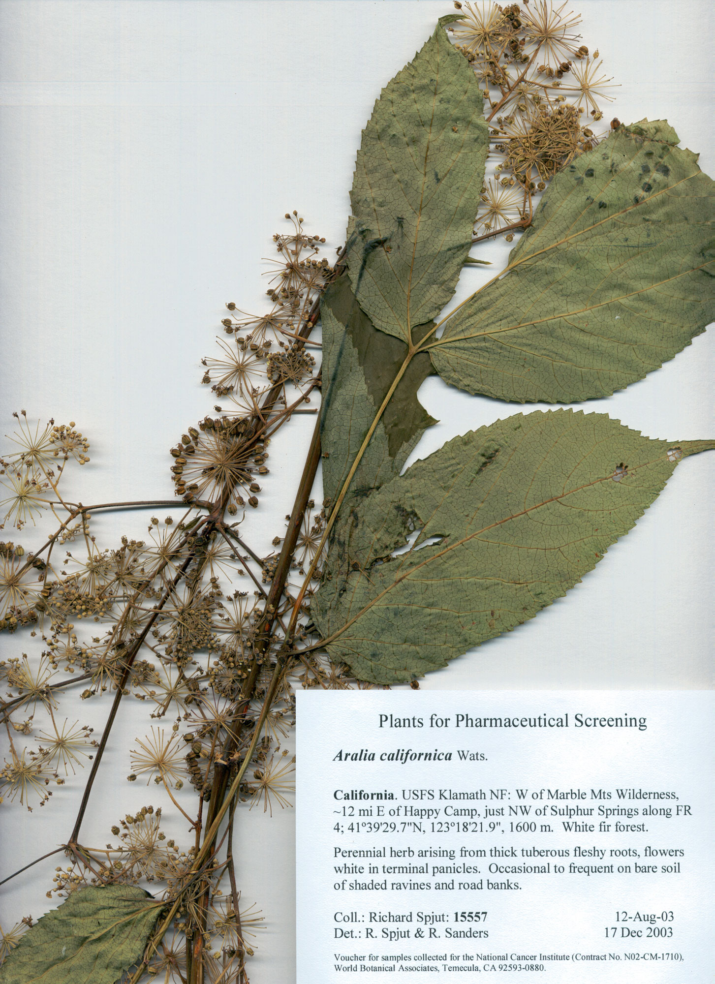
Aralia nudicaule—Historical collection data for cancer research |
|
Baek Y. H., J. E. Huh, J. D. Lee, D. Y. Choi and D. S. Park. 2006. Effect of Aralia cordata extracts on cartilage protection and apoptosis inhibition. Biol. Pharm. Bull. 2006 29(7): 1423–1430. “Cartilage loss in osteoarthritis is characterized by cartilage degradation and chondrocyte death. Cartilage degradation is induced by activation of matrix-metalloproteinases (MMPs) activity and degradation of glycosaminoglycan (GAG) and collagen. Also, chondrocyte death is induced by the apoptosis through the activation of MAP kinase and caspases activities. On the basis of this background, our study was designed to examine the cartilage protective and anti-apoptotic effect of Aralia cordata. Cartilage explants and Chondrocytes were cultured from rabbit knee joint cartilage and treated by 5 ng/ml IL-1alpha. Cartilage and chondroprotective effects of Aralia cordata were determined by measuring (1) GAG and collagen expression, (2) GAG and collagen degradation, (3) TIMP and MMPs expression, and (4) TIMP and MMPs activity. Anti-apoptotic effects of Aralia cordata were determined by measuring (1) JNK and p38 MAP kinase expression, (2) apoptotic cells by flow cytometry, and (3) caspase-3 activity. In cartilage explants and chondroctyes treated by IL-1alpha, Aralia cordata showed the decrease of GAG and collagen degradation, decrease of MMPs (MMP-1, -3, -13) activity, and increase of TIMP-1 activity in a dose-dependent manner. Aralia cordata also showed anti-apoptotic effect by inhibition of early and late apoptotic cells, sub-G1 phase cells, and caspase-3 activity through the downregulation of JNK and p38 MAP kinase signaling pathway. Aralia cordata inhibited the cartilage and chondrocyte destruction through the downregulation of MMPs activities and the inhibition of proteoglycan and collagen degradation. Also, Aralia cordata inhibited the chondrocyte apoptosis through the downregulation of JNK and p38 MAP kinase signal, and the inhibition of caspase-3 activity.” Bhat ZA, S. H. Ansari, H. M. Mukhtar, T. Naved, J. I. Siddiqui and N. A. Khan. 2005. Effect of Aralia cachemirica Decne root extracts on blood glucose level in normal and glucose loaded rats. Pharmazie 60(9): 712–713. “An aqueous and alcoholic extract of the roots of Aralia cachemirica (Araliaceae) were evaluated for hypoglycemic activity in normal fasted and glucose induced hyperglycemic rats. The aqueous and alcoholic extracts at a dose of 250 mg/kg showed statistically significant (p < 0.01) hypoglycemic activity in glucose loaded animals however no effect was observed in normal fasted rats.”
Chung Y.S., Y.
H. Choi, S. J. Lee, S. A. Choi, J. H. Lee, H. Kim and E. K. Hong.
2005. Water extract of Aralia elata prevents cataractogenesis in
vitro and in vivo. J. Ethnopharmacol. 2005 Oct 3;101(1-3):49-54. “The
water extract of Aralia elata (Aralia extract) has been used
in Korean traditional medicine to treat diabetes mellitus. Here, we
investigated the aldose reductase inhibitory activity, antioxidant
activity and anticataract capacity of Aralia extract using various
experimental systems. To determine its aldose reductase inhibitory
activity and its antioxidant effect, we used rat lens homogenates. Rat
lens cultures and a rat model were used to observe anticataract activity.
The resulting IC50 value of Aralia extract in vitro as an aldose reductase
inhibitor was 11.3 microg/ml and as an antioxidant was 25.1 microg/ml. Rat
lenses in media containing 20 mM xylose developed a distinctly opaque ring
in 9h, and treatment with Aralia extract at a concentration of 1mg/ml
lowered lens opacity by 36.4 and 31.3% after 24 and 48 h, respectively. In
vivo experiments were performed with streptozotocin (STZ) induced diabetic
rats. The diabetic control animals developed cataracts at 11 weeks after
STZ injection, while oral Aralia extract administered at 300 and 600 mg/kg
body weight for 11 weeks reduced cataract formation by 15 and 12%,
respectively. The present study shows that Aralia extract inhibits
aldose reductase and acts in vitro as an antioxidant, and suggests that
these activities have a preventive effect on cataractogenesis in xylose
containing lens organ cultures and in in vivo in STZ induced diabetic
rats.”
Jeong S. I.,
W. S. Han, Y. H. Yun and K. J. Kim. 2006. Continentalic acid from
Aralia continentalis shows activity against methicillin-resistant
Staphylococcus aureus. Lee E. B., O. J. Kim, S. S. Kang and C. Jeong. 2005. Araloside A, an antiulcer constituent from the root bark of Aralia elata. Biol. Pharm. Bull. 28(3): 523–526. “Araloside A, a potent inhibitor of gastric lesion and ulcer formation in rats, was isolated from the root bark of Aralia elata through a bioassay-guided separation procedure. The compound exhibited significant reduction of HCl.ethanol-induced gastric lesions and aspirin-induced gastric ulcers at oral doses of 50 and 100 mg/kg, respectively. These activities are comparable to those of cimetidine.” Tian Z, G. Lin, R. X. Zheng, F. Huang, M. S. Yang and P. G. Xiao. 2006. Anti-hepatoma activity and mechanism of ursolic acid and its derivatives isolated from Aralia decaisneana. World J. Gastroenterol. 12(6): 874–879. “AIM: To investigate the anti-tumor activity of ursolic acid (UA) and its derivatives isolated from Aralia decaisneana on hepatocellular carcinoma both in vitro and in vivo. METHODS: In vivo cytotoxicity was first screened by 3-[4,5-dimethylthiazol-2-yl]-2, 5-diphenyltetrazolium bromide (MTT) assay. Morphological observation, DNA ladder, flow cytometry analysis, Western blot and real time PCR were employed to elucidate the cytotoxic mechanism of UA. Implanted mouse hepatoma H22 was used to evaluate the growth inhibitory effect of UA in vivo. RESULTS: UA could significantly inhibit the proliferation of HepG2 and its drug-resistance strain, R-HepG2 cells, but had no inhibitory effect on primarily cultured normal mouse hepatocytes whereas all the six derivatives of UA could not inhibit the growth of all tested cell lines. Further study on mechanism demonstrated that apoptosis and G0/G1 arrest were involved in the cytotoxicity and cleavage of poly-(ADP-ribose)-polymerase (PARP). Downregulation of cyclooxygenase-2 (COX-2) protein and upregulation of heat shock protein (HSP) 105 mRNA correlated to the apoptosis of HepG2 cells treated with UA. In addition, UA also could inhibit the growth of H22 hepatoma in vivo. CONCLUSION: UA is a promising anti-tumor agent, but further work needs to be done to improve its solubility.” Tomatsu M., M. Ohnishi-Kameyama and N. Shibamoto. 2003. Aralin, a new cytotoxic protein from Aralia elata, inducing apoptosis in human cancer cells. Cancer Lett. 199(1): 19–25. “In this study, we purified a novel cytotoxic protein, aralin, from the shoots of Aralia elata. Aralin is composed of two subunits, A and B chains whose molecular weights are 29,100 and 32,200, respectively. In the assay using a normal human lung fibroblast cells (WI-38) and its SV40-transformed cells (VA-13), aralin demonstrated selective cytotoxicity against the virus-transformed cell line; the IC50 values of WI-38 and VA-13 were 10 and 0.8 ng/ml, respectively. Aralin showed positive response to DNA fragmentation in human lymphocyte HL-60 cells, and caspase specific inhibitors suppressed aralin-induced DNA fragmentation. These results indicate that the cytotoxicity of aralin is brought about primarily through the induction of apoptosis. Aralin also exhibited potent cytotoxic activity against various types of human cancer cell lines; cervical carcinoma cells (HeLa) proved the most sensitive, with an IC50 value of 0.08 ng/ml.” Wang J., Q. Li, G. Ivanochko and Y. Huang. 2006. Anticancer effect of extracts from a North American medicinal plant--wild sarsaparilla. Anticancer Res. 26(3A): 2157–2164. “The wild sarsaparilla (Aralia nudicaulis) plant is richly distributed in North America, mainly in Canada. In the present study, 24 extracts were obtained from the rhizome, stem, leaf and fruit of wild sarsaparilla. In the presence of RH (hexane fraction from the rhizome), the survival rate of WiDr (human colon cancer cell) was 3.5 +/- 2.7% (IC50 = 30.1 +/- 3.5 microg/ml) and that of Molt (human leukemia cell) was 2.4 +/- 2.8% (IC50 = 7.0 +/- 0.6 microg/ml). The survival rate of HELA (human cervix cancer cell) was only 1.8 +/- 0.9% in the presence of FH (hexane fraction from the fruit of wild sarsaparilla) (IC50 = 33.3 +/- 2.7 microg/ml). The cytotoxicities of RH and FH against normal human umbilical vein endothelial cells were significantly lower than against the tested human cancer cells. RH appeared to be the best extract against WiDr and Molt, whereas FH was the most effective against HELA. Because of the rich natural supply, simple extraction procedure and high yield, RH and FH of wild sarsaparilla have the potential to be developed into selective anticancer nutraceutical and/or pharmaceutical products with few side-effects and low cost.” |
|